Learn More
Invitrogen™ CD279 (PD-1) Monoclonal Antibody (J43), Brilliant Violet™ 605, eBioscience™
Descripción
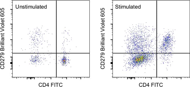
Description The J43 monoclonal antibody reacts with mouse PD-1 (programmed death-1), a 55 kDa member of the Ig superfamily. It is reported that J43 inhibits the binding of mouse PD-L1-Ig and mouse PD-L2-Ig to PD-1/BHK transfected cells. When administrated in vivo, both intact and Fab of J43 are reported to enhance contact hypersensitivity and exacerbate acute GVHD similar to transfer of PD-1-deficient cells. Injection of J43 also exacerbates EAE and NOD diabetes as do specific antibodies to mouse PD-L1 and PD-L2. Applications Tested This J43 antibody has been tested by flow cytometric analysis of mouse splenocytes. This may be used at less than or equal to 0.5 μg per test. A test is defined as the amount (μg) of antibody that will stain a cell sample in a final volume of 100 μL. Cell number should be determined empirically but can range from 10^5 to 10^8 cells/test. It is recommended that the antibody be carefully titrated for optimal performance in the assay of interest. Blocking Buffers When using two or more Super Bright, Brilliant Violet™, Brilliant Ultra Violet™, or other polymer dye-conjugated antibodies in a staining panel, it is recommended to use Super Bright Complete Staining Buffer (Product # SB-4401 ) or Brilliant Stain Buffer (Product # 00-4409-75 ) to minimize any non-specific polymer interactions. Please refer to the datasheet for Super Bright Staining Buf...
Especificaciones
Especificaciones
| Antígeno | CD279 (PD-1) |
| Aplicaciones | Flow Cytometry |
| Clasificación | Monoclonal |
| Clon | J43 |
| Concentración | 0.2 mg/mL |
| Conjugado | Brilliant Violet 605 |
| Formulación | PBS with BSA and 0.09% sodium azide; pH 7.2 |
| génica | Pdcd1 |
| N.º de referencia del gen | Q02242 |
| Alias de gen | CD279; EGK_05005; hPD1; hPD-1; hPD-l; hSLE1; Ly101; mPD-1; PD1; PD-1; Pdc1; Pdcd1; programmed cell death 1; programmed cell death 1 protein; programmed cell death protein 1; programmed cell death protein 1-like; programmed death 1; Protein PD1; protein PD-1; sCD279; SLEB2; soluble CD279; systemic lupus erythematosus susceptibility 2 |
| Mostrar más |
Al hacer clic en Enviar, acepta que Fisher Scientific se ponga en contacto con usted en relación con los comentarios que ha proporcionado en este formulario. No compartiremos su información para ningún otro fin. Toda la información de contacto proporcionada se mantendrá de acuerdo con nuestra Política de Privacidad. Política de privacidad.